Source:- Google.com.pk
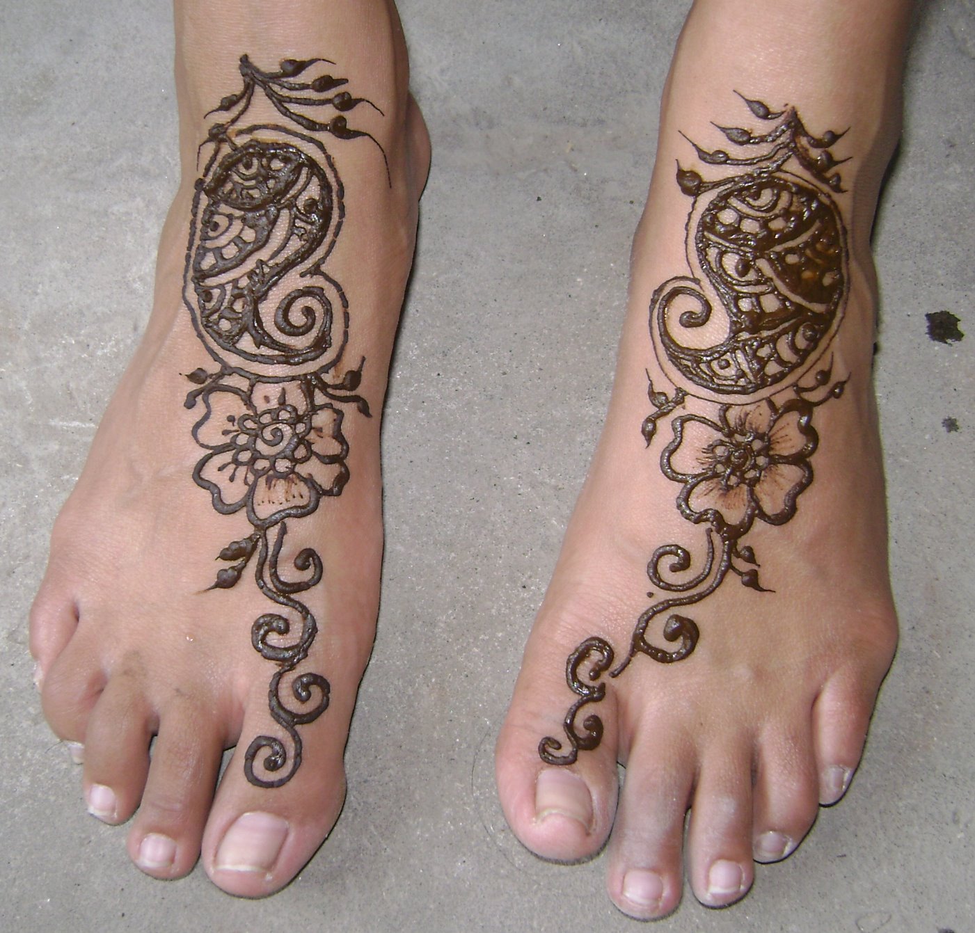
The foot (plural feet) is an anatomical structure found in many vertebrates. It is the terminal portion of a limb which bears weight and allows locomotion. In many animals with feet, the foot is a separate organ at the terminal part of the leg made up of one or more segments or bones, generally including claws or nails.The human foot and ankle is a strong and complex mechanical structure containing 26 bones, 33 joints (20 of which are actively articulated), and more than a hundred muscles, tendons, and ligaments.
An anthropometric study of 1197 North American adult Caucasian males (mean age 35.5 years) found that a man's foot length was 26.3 cm with a standard deviation of 1.2 cm.The foot can be subdivided into the hindfoot, the midfoot, and the forefoot:The hindfoot is composed of the talus (or ankle bone) and the calcaneus (or heel bone). The two long bones of the lower leg, the tibia and fibula, are connected to the top of the talus to form the ankle. Connected to the talus at the subtalar joint, the calcaneus, the largest bone of the foot, is cushioned inferiorly by a layer of fat.The five irregular bones of the midfoot, the cuboid, navicular, and three cuneiform bones, form the arches of the foot which serves as a shock absorber. The midfoot is connected to the hind- and fore-foot by muscles and the plantar fascia.The forefoot is composed of five toes and the corresponding five proximal long bones forming the metatarsus. Similar to the fingers of the hand, the bones of the toes are called phalanges and the big toe has two phalanges while the other four toes have three phalanges. The joints between the phalanges are called interphalangeal and those between the metatarsus and phalanges are called metatarsophalangeal (MTP).Both the midfoot and forefoot constitute the dorsum (the area facing upwards while standing) and the planum (the area facing downwards while standing).
The instep is the arched part of the top of the foot between the toes and the ankle.The feet of a newborn infant.
tibia, fibula tarsus: talus, calcaneus, cuneiformes, cuboid, and navicular
metatarsus: first, second, third, fourth, and fifth metatarsal bone
phalanges
There can be many sesamoid bones near the metatarsophalangeal joints, although they are only regularly present in the distal portion of the first metatarsal bone.Arches of the footThe human foot has two longitudinal arches and a transverse arch maintained by the interlocking shapes of the foot bones, strong ligaments, and pulling muscles during activity. The slight mobility of these arches when weight is applied to and removed from the foot makes walking and running more economical in terms of energy. As can be examined in a footprint, the medial longitudinal arch curves above the ground. This arch stretches from the heel bone over the "keystone" ankle bone to the three medial metatarsals. In contrast, the lateral longitudinal arch is very low. With the cuboid serving as its keystone, it redistributes part of the weight to the calcaneus and the distal end of the fifth metatarsal. The two longitudinal arches serve as pillars for the transverse arch which run obliquely across the tarsometatarsal joints. Excessive strain on the tendons and ligaments of the feet can result in fallen arches or flat feet.The muscles acting on the foot can be classified into extrinsic muscles, those originating on the anterior or posterior aspect of the lower leg, and intrinsic muscles, originating on the dorsal (top) or plantar (base) aspects of the foot.All muscles originating on the lower leg except the popliteus muscle are attached to the bones of the foot. The tibia and fibula and the interosseous membrane separate these muscles into anterior and posterior groups, in their turn subdivided into subgroups and layers.Extensor group: tibialis anterior originates on the proximal half of the tibia and the interosseous membrane and is inserted near the tarsometatarsal joint of the first digit. In the non-weight-bearing leg tibialis anterior flexes the foot dorsally and lift its medial edge (supination). In the weight-bearing leg it brings the leg towards the back of the foot, like in rapid walking. Extensor digitorum longus arises on the lateral tibial condyle and along the fibula to be inserted on the second to fifth digits and proximally on the fifth metatarsal. The extensor digitorum longus acts similar to the tibialis anterior except that it also dorsiflexes the digits. Extensor hallucis longus originates medially on the fibula and is inserted on the first digit. As the name implies it dorsiflexes the big toe and also acts on the ankle in the unstressed leg. In the weight-bearing leg it acts similar to the tibialis anterior.
Peroneal group: peroneus longus arises on the proximal aspect of the fibula and peroneus brevis below it on the same bone. Together, their tendons pass behind the lateral malleolus. Distally, peroneus longus crosses the plantar side of the foot to reach its insertion on the first tarsometatarsal joint, while peroneus brevis reaches the proximal part of the fifth metatarsal. These two muscles are the strongest pronators and aid in plantar flexion. Longus also acts like a bowstring that braces the transverse arch of the foot.The superficial layer of posterior leg muscles is formed by the triceps surae and the plantaris. The triceps surae consists of the soleus and the two heads of the gastrocnemius. The heads of gastrocnemius arise on the femur, proximal to the condyles, and soleus arises on the proximal dorsal parts of the tibia and fibula. The tendons of these muscles merge to be inserted onto the calcaneus as the Achilles tendon. Plantaris originates on the femur proximal to the lateral head of the gastrocnemius and its long tendon is embedded medially into the Achilles tendon. The triceps surae is the primary plantar flexor and its strength becomes most obvious during ballet dancing. It is fully activated only with the knee extended because the gastrocnemius is shortened during knee flexion. During walking it not only lifts the heel, but also flexes the knee, assisted by the plantaris.the deep layer of posterior muscles tibialis posterior arises proximally on the back of the interosseous membrane and adjoining bones and divides into two parts in the sole of the foot to attach to the tarsus. In the non-weight-bearing leg, it produces plantar flexion and supination, and, in the weight-bearing leg, it proximates the heel to the calf. flexor hallucis longus arises on the back of the fibula (i.e. on the lateral side), and its relatively thick muscle belly extends distally down to the flexor retinaculum where it passes over to the medial side to stretch across the sole to the distal phalanx of the first digit. The popliteus is also part of this group, but, with its oblique course across the back of the knee, does not act on the foot.On the back (top) of the foot, the tendons of extensor digitorum brevis and extensor hallucis brevis lie deep to the system of long extrinsic extensor tendons. They both arise on the calcaneus and extend into the dorsal aponeurosis of digits one to four, just beyond the penultimate joints. They act to dorsiflex the digits.
Plantar aspects of foot, varying depths (superficial to deep)Similar to the intrinsic muscles of the hand, there are three groups of muscles in the sole of foot, those of the first and last digits, and a central group:Muscles of the big toe: abductor hallucis stretches medially along the border of the sole, from the calcaneus to the first digit. Below its tendon, the tendons of the long flexors pass through the tarsal canal. It is an abductor and a weak flexor, and also helps maintain the arch of the foot. flexor hallucis brevis arises on the medial cuneiform bone and related ligaments and tendons. An important plantar flexor, it is crucial for ballet dancing. Both these muscles are inserted with two heads proximally and distally to the first metatarsophalangeal joint. Adductor hallucis is part of this group, though it originally formed a separate system (see contrahens.) It has two heads, the oblique head originating obliquely across the central part of the midfoot, and the transverse head originating near the metatarsophalangeal joints of digits five to three. Both heads are inserted into the lateral sesamoid bone of the first digit. Adductor hallucis acts as a tensor of the plantar arches and also adducts the big toe and then might plantar flex the proximal phalanx.Muscles of the little toe: Stretching laterally from the calcaneus to the proximal phalanx of the fifth digit, abductor digiti minimi form the lateral margin of the foot and is the largest of the muscles of the fifth digit. Arising from the base of the fifth metatarsal, flexor digiti minimi is inserted together with abductor on the first phalanx. Often absent, opponens digiti minimi originates near the cuboid bone and is inserted on the fifth metatarsal bone. These three muscles act to support the arch of the foot and to plantar flex the fifth digit.
Pakistani Girls Feet Hot Pakistani Girls Mobile Numbers Names Hair Styles Images Funny Pics Photos
Pakistani Girls Feet Hot Pakistani Girls Mobile Numbers Names Hair Styles Images Funny Pics Photos
Pakistani Girls Feet Hot Pakistani Girls Mobile Numbers Names Hair Styles Images Funny Pics Photos
Pakistani Girls Feet Hot Pakistani Girls Mobile Numbers Names Hair Styles Images Funny Pics Photos
Pakistani Girls Feet Hot Pakistani Girls Mobile Numbers Names Hair Styles Images Funny Pics Photos
Pakistani Girls Feet Hot Pakistani Girls Mobile Numbers Names Hair Styles Images Funny Pics Photos
Pakistani Girls Feet Hot Pakistani Girls Mobile Numbers Names Hair Styles Images Funny Pics Photos
Pakistani Girls Feet Hot Pakistani Girls Mobile Numbers Names Hair Styles Images Funny Pics Photos
Pakistani Girls Feet Hot Pakistani Girls Mobile Numbers Names Hair Styles Images Funny Pics Photos
Pakistani Girls Feet Hot Pakistani Girls Mobile Numbers Names Hair Styles Images Funny Pics Photos
Pakistani Girls Feet Hot Pakistani Girls Mobile Numbers Names Hair Styles Images Funny Pics Photos


No comments:
Post a Comment